Salivary Gland Disorders
Salivary Gland Stones
Salivary Stones are uncommon. The incidence in the UK population is about 60 cases per million population each year. The majority (80%) form in the submandibular gland (SMG) and 20% in the Parotid. A myth has developed that once a stone forms, the gland becomes infected and never recovers. We have shown this is not the case but most surgical units will recommend gland removal. This is no longer necessary. Salivary stones can be reliably removed and the gland remains healthy.
The management of salivary stones was revolutionised by teams in London, Paris, Erlangen, Israel and Milan. We published the combined results from all these centres which totalled more than 4,600 patients with salivary stones. Less than 3% of patients required salivary gland removal with 82% of stones retrieved. The results of stone removal today (2017) are an improvement on this figure with over 90% of stones extracted by minimally invasive techniques.
Techniques available
New miniature instruments have made minimally invasive salivary gland surgery possible. New micro-endoscopes are less than 1mm in diameter and allow excellent visualisation of the salivary duct system. Micro-instruments such as baskets, balloons, forceps and lithotripter wires can be passed through the endoscopes to target stones and strictures.
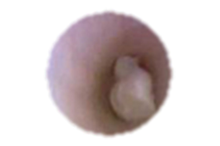

Small stones
The salivary ducts are 2 to 3 mm wide. Small stones less than 4mm can be grasped and pulled down the duct. This can be done with tiny wire baskets either under direct vision through a micro-endoscope or under radiological control. The results are excellent with about 75% of small stones retrieved by this method alone.
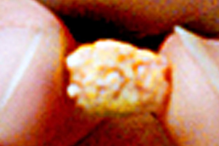
Minimum sized stones
Stones measuring between 5 and 8 mm cannot be drawn down the duct. They have to be broken up first. A new lithotripter (stone breaker) is now available, it is hand-held and works by passing a thin wire down the working channel of the micro endoscope to touch the stone. A shock wave is passed down the wire and after approximately 50 shocks the stone will break and the fragments can be removed with a basket. (video bottom right) If cases are selected appropriately then in our experience about 75% of middle size stones can be successfully removed by this method.
Parotid gland stones larger than 8mm
These larger stones can be removed more easily and efficiently by minimally invasive surgical procedures that leave the gland intact.
These stones are located with the endoscope light and camera and illuminated from the inside to guide surgery – video below.
We have published our results in over 100 cases with excellent rates of stone retrieval (95%+) and few serious complications.
An endoscope-mounted camera with light can accurately locate the stone within the duct and guide the surgery.
Video: Endoscopically assisted stone removal from parotid gland
Fixed submandibular stones larger than 8mm
These are also removed by an endoscope assisted surgical technique. Under day care general anaesthesia, an incision in made along the floor of the mouth, the submandibular duct is found and traced back into the gland with the help of the micro endoscope. The stone is found and released. The operation takes about 30-40min. Our results in over 300 cases show excellent results with stone retrieval rates of 97%.
Other available channels in the endoscope carry light, camera, fibre-optics and other devices.