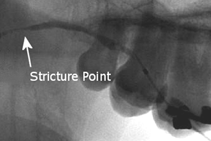
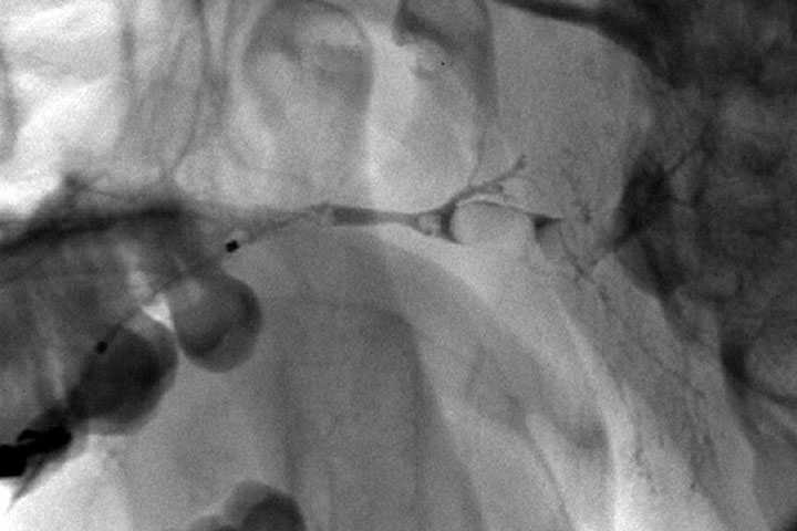
A stricture occurs when part of the salivary duct becomes narrowed or blocked through inflammation, pinching or scarring.
When the salivary gland is relatively inactive, saliva stagnates and forms a mucus plug behind the stricture. When saliva is produced at mealtime, the backlog of saliva behind the stricture causes swelling, and the increased pressure inside the gland causes pain.
If and when the mucus plug is dislodged by the pressure, the built up saliva is released into the mouth and the swelling subsides. This is accompanied by a bad taste in the mouth.
Traditionally there was no way of managing strictures apart from removing the salivary gland.
How common are they?
There are about 20 stricture cases per million annually. Salivary stone blockages and strictures present similar symptoms, about 1 in 4 patients with blockage symptoms (gland swells at mealtime) have a stricture in the duct wall. The majority of strictures are in the parotid (75%) More strictures occur in women than men.
The cause of strictures is unknown.
Stricture Types
Inflammation
When using the endoscope, sometimes the duct is very inflamed as if the patient were suffering an allergic reaction. It is thought that the inflammation leads to scar formation and strictures. These ducts can be washed with steroid solution to reduce the inflammation.
Point stricture
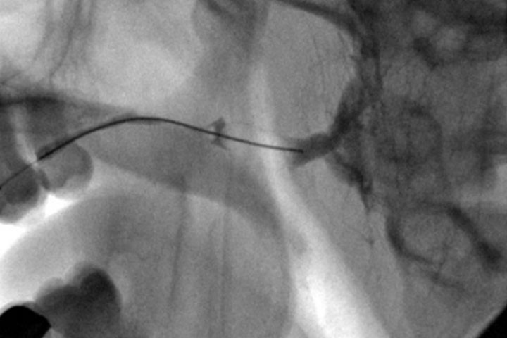
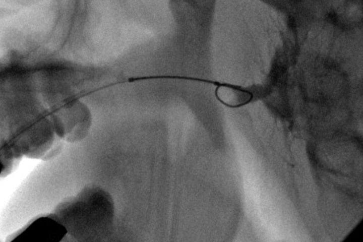
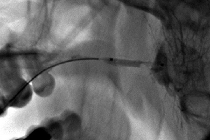
These strictures can be dilated with balloons or thin dilators. This is done under endoscopic or radiological control. The duct is washed with steroid solution at the end.
Diffuse stricture
On occasion the whole duct is scarred along its length. These ducts are more difficult to treat, but gradual dilation and washouts improve the symptoms.
Minimally Invasive Methods of Treating Salivary Strictures
New micro-endoscopes are less than 1mm in diameter and allow excellent visualisation of the salivary duct system. Micro-instruments such as baskets, drills and balloons can be passed through the endoscopes to target strictures.
There are two approaches, both provided in the outpatient setting under local anaesthesia
Endoscope assisted dilation of stricture
An endoscope is introduced into the salivary duct. Mild strictures can be dilated flushing the duct with a saline solution under pressure. Tighter strictures can be opened will a small hand drill or by a small balloon that can be inflated within the stricture.